 English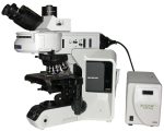
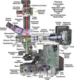
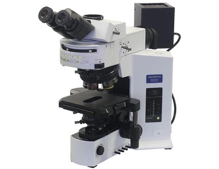
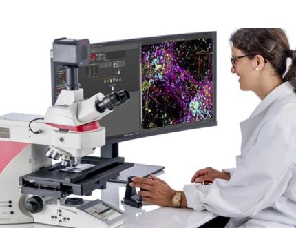
میکروسکوپ نوری / فلورسانس Olympus BX53
معرفی محصول
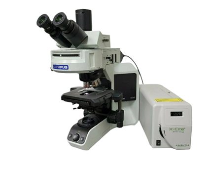
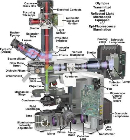
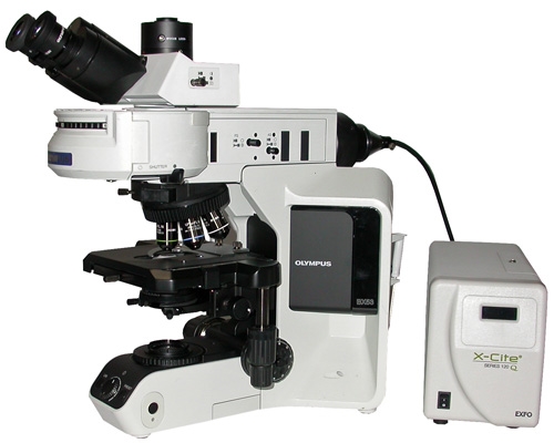
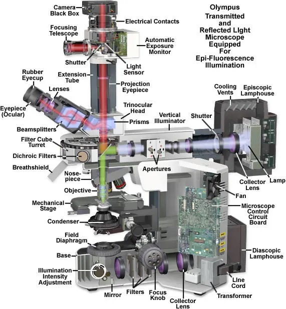
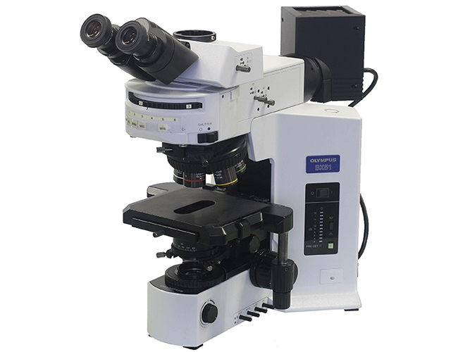
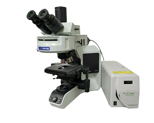
میکروسکوپ Olympus BX53 یک میکروسکوپ تحقیقاتی حرفهای و پیشرفته از برند معتبر المپوس است که برای مشاهده و تصویربرداری از نمونههای میکروسکوپی با استفاده از نور عبوری (Brightfield) و فلورسانس طراحی شده است.
این سیستم یک پلتفرم قدرتمند برای تحقیقات آزمایشگاهی محسوب میشود و بهطور گسترده در زیستشناسی سلولی و مولکولی، پاتولوژی، میکروبیولوژی، تحقیقات دارویی و همچنین آزمایشگاههای دانشگاهی و صنعتی مورد استفاده قرار میگیرد.
BX53 به کاربران این امکان را میدهد که ساختارهای طبیعی نمونه را با نور معمولی مشاهده کنند و همچنین اجزای سلولی یا مولکولی نشاندار شده با رنگهای فلورسنت را با وضوح و دقت بسیار بالا تصویربرداری نمایند.
طراحی ماژولار و قابل ارتقاء
یکی از مهمترین مزایای Olympus BX53 طراحی کاملاً ماژولار آن است.
این سیستم را میتوان متناسب با نیاز فعلی پیکربندی کرد و در هر زمان بدون نیاز به تعویض کل دستگاه ارتقاء داد.
BX53 میتواند هم بهعنوان یک میکروسکوپ آموزشی ساده و هم بهعنوان یک سیستم پیشرفته تصویربرداری فلورسانس مورد استفاده قرار گیرد، بسته به نوع کاربرد.
فیلترهای فلورسانس (کانالها)
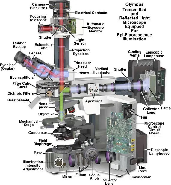
BX53 از چندین مکعب فیلتر فلورسانس بهصورت همزمان پشتیبانی میکند (معمولاً بین ۳ تا ۸ موقعیت در برجک فیلتر)، که امکان تصویربرداری چندرنگ را فراهم میسازد.
فیلترهای رایج عبارتاند از:
DAPI (آبی) – برای رنگآمیزی هسته سلول
FITC / GFP (سبز) – برای پروتئینها و رنگهای فلورسنت سبز
TRITC / Cy3 (نارنجی-قرمز)
Texas Red / mCherry
Cy5 / Alexa Fluor 647 (نزدیک مادون قرمز)
فیلترهای تخصصی دیگر مانند CFP، YFP و سری Alexa Fluor
🔹 این فیلترها امکان شناسایی همزمان چندین بیومارکر را در یک نمونه فراهم میکنند که برای تحقیقات پیشرفته سلولی و مولکولی بسیار حیاتی است.
منابع نوری
میکروسکوپ BX53 بسته به نوع کاربرد میتواند به منابع نوری مختلفی مجهز شود:
برای تصویربرداری نوری (Brightfield):
نور LED سفید با یکنواختی بالا و رنگ واقعی
یا لامپ هالوژن با شدت قابل تنظیم
برای تصویربرداری فلورسانس:
منابع نور متال هالید (مثل X-Cite 120W) با شدت بالا
لامپهای جیوه
لامپهای زنون
منابع LED فلورسانس نسل جدید با طول عمر بالا، گرمای کم و سوئیچ سریع
🔹 این انعطافپذیری باعث میشود تحریک فلورسانس در بازه طیفی گسترده از DAPI تا Cy5 با کنتراست و کیفیت بالا انجام شود.
ویژگیهای کلیدی فروش
1. تصویربرداری فلورسانس چندکاناله
امکان مشاهده و ثبت همزمان چند کانال فلورسانس از یک نمونه.
2. پلتفرم کاملاً قابل ارتقاء
قابل توسعه با:
فیلترهای فلورسانس بیشتر
منابع نوری پیشرفتهتر
دوربینهای حساس CCD یا CMOS
نرمافزارهای حرفهای تصویربرداری و تحلیل
3. اتصال به دوربین و کامپیوتر
سازگار با دوربینهای دیجیتال برای:
ثبت تصاویر با وضوح بالا
ضبط ویدئو
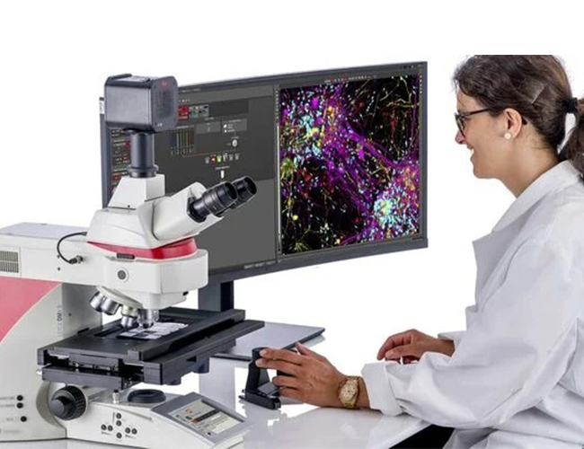
تحلیل کمی تصاویر با نرمافزارهایی مانند cellSens یا ImageJ
4. قابلیتهای موتوردار (اختیاری)
در پیکربندیهای پیشرفته:
استیج موتوردار برای اسکن خودکار
تصویربرداری Z-stack
تایملپس (Time-lapse)
اتصال تصاویر (Stitching) برای نمونههای بزرگ
5. کیفیت اپتیکی فوقالعاده
مجهز به اپتیکهای حرفهای Olympus با:
تفکیکپذیری بسیار بالا
کنتراست عالی
بازتولید دقیق و طبیعی رنگها
کاربردها
تحقیقات زیستشناسی سلولی و مولکولی
تصویربرداری فلورسانس چندرنگ
پاتولوژی و هیستولوژی
تحقیقات دارویی و پزشکی
آزمایشگاههای تحقیق و توسعه دانشگاهی و صنعتی