 English
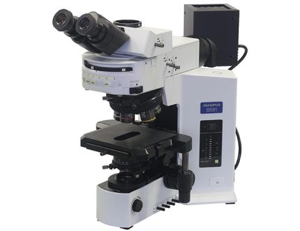
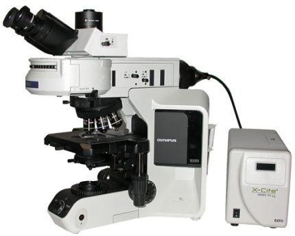
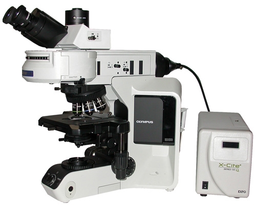
میکروسکوپ فلورسانس Olympus BX43
معرفی محصول
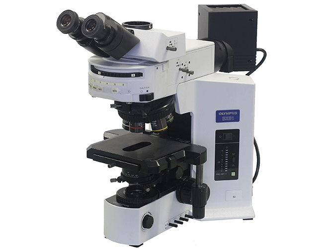
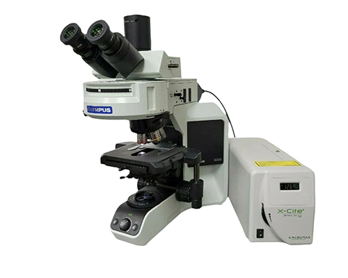
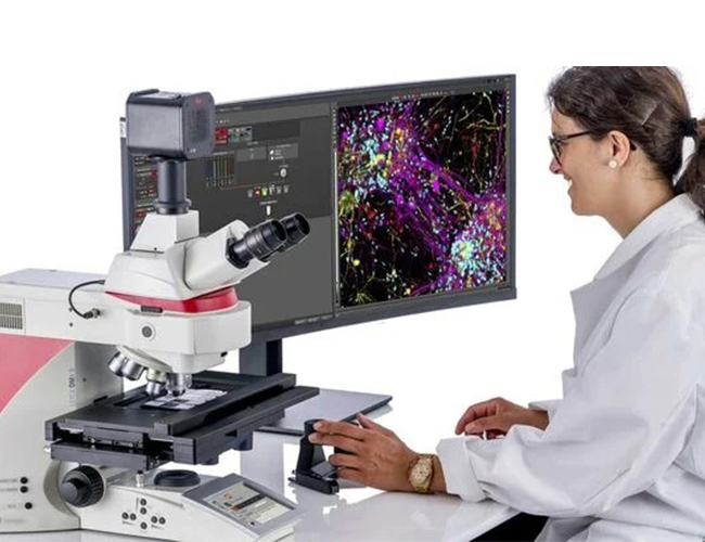
میکروسکوپ Olympus BX43 یک سیستم تحقیقاتی پیشرفته و با عملکرد بالا است که برای تصویربرداری با نور عبوری (Brightfield) و فلورسانس طراحی شده است. این مدل ترکیبی از کیفیت اپتیکی عالی، انعطافپذیری و قابلیت ارتقاء ماژولار است و در آزمایشگاههای تحقیقاتی، تشخیصی و آموزشی کاربرد فراوان دارد.
BX43 در حوزههای زیستشناسی سلولی و مولکولی، ژنتیک، پاتولوژی، سیتوژنتیک، میکروبیولوژی و تحقیقات پزشکی مورد استفاده قرار میگیرد.
این سیستم به کاربران امکان میدهد ساختارهای طبیعی سلولها را با نور معمولی مشاهده کنند و همچنین مولکولها و اجزای سلولی نشاندار شده با فلورسانس را با وضوح و دقت بالا شناسایی و تصویربرداری کنند.
طراحی ماژولار و حرفهای
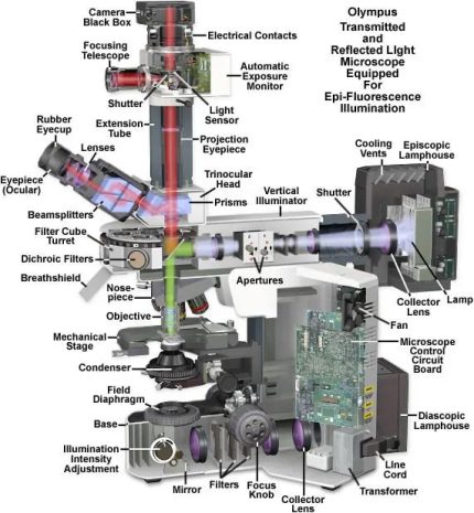
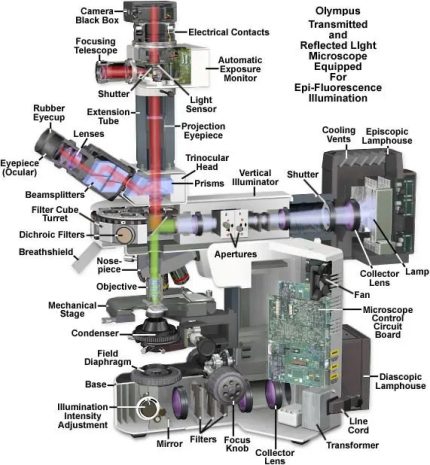
Olympus BX43 دارای طراحی ماژولار و انعطافپذیر است که امکان پیکربندی متناسب با نیازهای آزمایشگاه را فراهم میکند.
کاربران میتوانند بخشهای فلورسانس، فیلترها، منابع نوری، دوربینها و نرمافزارهای تصویربرداری را بدون نیاز به تعویض کل میکروسکوپ اضافه یا ارتقاء دهند.
BX43 برای آزمایشگاههایی که به دنبال ترکیب عملکرد حرفهای و قابلیت ارتقاء آسان هستند، گزینهای ایدهآل است.
فیلترهای فلورسانس (کانالها)
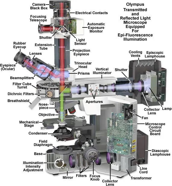
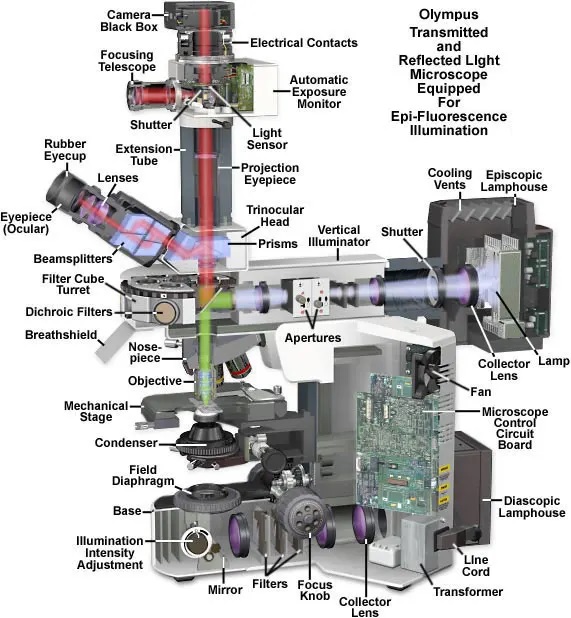
BX43 از چندین مکعب فیلتر فلورسانس (معمولاً ۳ تا ۶ موقعیت در برجک فیلتر) پشتیبانی میکند و امکان تصویربرداری چندکاناله را فراهم میسازد.
فیلترهای رایج شامل:
DAPI (آبی) – برای رنگآمیزی هسته
FITC / GFP (سبز)
TRITC / Cy3 (نارنجی-قرمز)
Texas Red / mCherry
Cy5 / Alexa Fluor 647
فیلترهای اختیاری CFP و YFP
🔹 این فیلترها امکان شناسایی همزمان چندین بیومارکر در یک نمونه را فراهم میکنند.
منابع نوری
BX43 میتواند به منابع نوری مختلف مجهز شود:
برای تصویربرداری نوری (Brightfield):
لامپ هالوژن با شدت قابل تنظیم
نور LED با رنگ طبیعی و یکنواخت
برای تصویربرداری فلورسانس:
لامپ جیوه (Mercury Lamp)
منابع نور متال هالید
منابع نور LED فلورسانس نسل جدید
این منابع نوری امکان تحریک فلورسانس مؤثر در طیف گسترده از DAPI تا Cy5 را فراهم میکنند.
کاربرد ویژه: FISH (فلورسانس در جایگاه هیبریدیزاسیون)
میکروسکوپ BX43 برای تکنیک FISH (Fluorescence In Situ Hybridization) مناسب است.
در روش FISH، از پروبهای فلورسنت برای شناسایی و مکانیابی توالیهای خاص DNA یا RNA در سلولها یا کروموزومها استفاده میشود.
ویژگیهایی که BX43 را برای FISH ایدهآل میکنند:
اپتیک با وضوح و دقت بالا
نور فلورسانس پایدار و یکنواخت
فیلترهای دقیق فلورسانس
امکان تصویربرداری چندکاناله
با BX43 میتوان به راحتی آزمایشهای FISH را برای مواردی مانند:
تحلیل سیتوژنتیک
شناسایی ناهنجاریهای کروموزومی
تشخیص سرطان و جهشهای ژنتیکی
بررسی ناباروری و IVF
انجام داد.
ویژگیهای کلیدی
1. تصویربرداری فلورسانس چندکاناله
امکان مشاهده همزمان چند پروب فلورسنت در یک نمونه.
2. اتصال به دوربین و نرمافزار
سازگار با دوربینهای CCD و CMOS برای:
ثبت تصاویر با وضوح بالا
مستندسازی نتایج FISH
تحلیل کمی تصاویر با نرمافزارهایی مانند ImageJ و cellSens
3. کیفیت اپتیکی حرفهای
لنزهای Olympus با:
وضوح بسیار بالا
کنتراست عالی
بازتولید طبیعی رنگها
4. قابلیت ارتقاء و انعطافپذیری
امکان افزودن:
فیلترهای فلورسانس بیشتر
منابع نوری LED یا متال هالید پیشرفته
سیستمهای تصویربرداری دیجیتال
کاربردها
تکنیک FISH و سیتوژنتیک
زیستشناسی سلولی و مولکولی
ژنتیک پزشکی
پاتولوژی و هیستولوژی
تحقیقات سرطان
آزمایشگاههای تحقیقاتی، تشخیصی و آموزشی